A High-Definition Surgical Atlas
Redefining Neuroanatomical Education
Where authentic human anatomy meets cutting-edge technology
The Origin
Why This Project Was Born
Anatomy is the irreplaceable foundation of every surgical act, especially in neurosurgery, where millimetric dissection of critical structures occurs in deep and confined spaces often distorted by pathology.
While anatomical lab access is expanding globally, daily hands-on dissection remains logistically challenging for many neurosurgical trainees. Limited lab time, institutional constraints, and geographic disparities still restrict consistent exposure to real human anatomy.
This atlas was created to:
- Provide the most realistic representation of human neuroanatomy
- Surpass the didactic limitations of synthetic or plastic models
- Maintain the scientific rigor of classical anatomical dissection
An Atlas Built by Neurosurgeons, for Neurosurgeons
This platform is designed around the real needs of neurosurgical training and operative planning. Every structure, corridor, and dissection step is presented with a surgical mindset.
The Method
Our Approach
Augmented Reality, Not Artificial Reality
Plastic models and synthetic reconstructions, while useful in early training, inevitably simplify and distort complex anatomy.
In contrast, the images and models within this atlas are derived exclusively from human anatomy.
The result is not a simulation, but a translation of true human anatomy into an interactive, explorable, and clinically useful format.
A Complement, Not a Substitute
Cadaveric dissection remains, and must remain, the gold standard for surgical anatomical training.
However, the ability to explore surgical corridors, observe progressive exposure of critical structures, and mentally simulate complex approaches, anywhere and at any time, makes this platform a powerful adjunct to laboratory-based training.
Our Mission
To offer neurosurgeons and trainees a scientifically rigorous, technologically advanced, and pedagogically effective tool that bridges the gap between theoretical anatomy and operative reality.
The People
Meet the Team
Two people at the intersection of medicine and technology, building the atlas that neurosurgeons deserve.
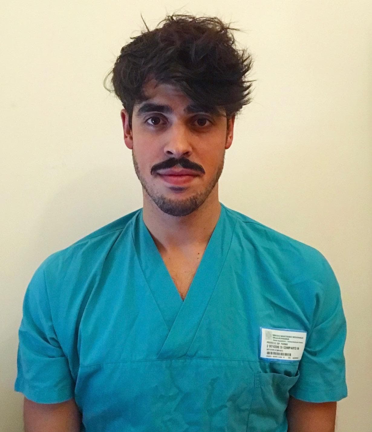
Dr. Luca Speranzon
- Medical doctor - University of Parma
- Resident in Neurosurgery - University of Catania
- Skull Base Microneurosurgery Former Fellow — Weill Cornell Medicine / NewYork-Presbyterian Hospital
- Harvard Medical School — Master in AI and Healthcare: From Strategies to Implementation
Alexandru Talpau
Partners
In Collaboration With
Partnering with leading academic and medical institutions.
Università di Catania
Weill Cornell Medicine
NewYork-Presbyterian Hospital
Bernardo & Evins Skull Base Neurosurgery
Ready to Explore?
Experience the future of neuroanatomical education with our interactive atlas.